
Fil:MitochondrionCAM.jpg
Ingen høyere oppløsning tilgjengelig.
MitochondrionCAM.jpg (618 × 409 piksler, filstørrelse: 37 KB, MIME-type: image/jpeg)
| Denne filen er fra Wikimedia Commons og kan brukes av andre prosjekter. Informasjonen fra filbeskrivelsessiden vises nedenfor. |
Beskrivelse
| Beskrivelse |
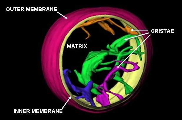
English: This is an image of a frozen-hydrated rat liver mitochondrion based on work done at the Wadsworth Center's Resource for Visualization of Biological Complexity (http://www.wadsworth.org/databank/electron/cryomito_dis2.html). It has not been previously published and I am releasing it into public domain for this article. The point is that the cristae are not folds of the inner membrane of the mitochondrion but pleomorphic invaginations with narrow tubular connections to each other and to the peripheral region of the inner membrane. I can advise on how to change your cartoon to be more accurate, while still retaining the basic format. There are numerous published references in last 10 years, the most recent: C.A. Mannella (2006) Biophysica et Biochimica Acta 1762: 140-147 |
| Dato | 17. juli 2006 (original upload date) |
| Kilde | Transferred from en.wikipedia to Commons by Gustavocarra. |
| Opphavsperson | Original uploader was Carmmann (talk) at en.wikipedia |
| Andre versjoner |
[]
|


.jpg)


Lisensiering
| Dette verket har blitt frigitt til allmennheten av opphavspersonen Carmmann at engelsk Wikipedia. Dette gjelder på verdensbasis. I enkelte land kan dette være juridisk umulig. I så fall: Carmmann gir hvem som helst retten til å bruke dette verket for ethvert formål, uten noen vilkår, med mindre slike vilkår kreves ved lov. |
Orginal opplastningslogg
The original description page was here. All following user names refer to en.wikipedia.
- 2006-07-17 02:14 Pschemp 618×409× (37898 bytes) cropped version
- 2006-02-05 02:40 Carmmann 720×540× (28251 bytes) This is an image of a frozen-hydrated rat liver mitochondrion based on work done at the Wadsworth Center's Resource for Visualization of Biological Complexity (http://www.wadsworth.org/databank/electron/cryomito_dis2.html). It has not been previously pub
Filhistorikk
Klikk på et tidspunkt for å vise filen slik den var på det tidspunktet.
| Dato/klokkeslett | Miniatyrbilde | Dimensjoner | Bruker | Kommentar | |
|---|---|---|---|---|---|
| nåværende | 15. aug. 2009 kl. 18:46 | | 618 × 409 (37 KB) | Gustavocarra | {{Information |Description={{en|This is an image of a frozen-hydrated rat liver mitochondrion based on work done at the Wadsworth Center's Resource for Visualization of Biological Complexity (http://www.wadsworth.org/databank/electron/cryomito_dis2.html). |
Filbruk
Det er ingen sider som bruker denne filen.
Global filbruk
Følgende andre wikier bruker denne filen:
- Bruk i ar.wikipedia.org
- Bruk i bs.wikipedia.org
- Bruk i cs.wikipedia.org
- Bruk i da.wikipedia.org
- Bruk i en.wikipedia.org
- Bruk i gl.wikipedia.org
- Bruk i kn.wikipedia.org
- Bruk i ml.wikipedia.org
- Bruk i uk.wikipedia.org